Find Doctors
Middle-aged man experiences rapid vision loss: urgent medical issue.
August 19 2022 | Medicover Hospitals | HyderabadTA carotid-cavernous fistula (CCF) results from an abnormal vascular shunt from the carotid artery to the venous channels of the cavernous sinus. The clinical features depend on the neurovascular structures involved in shunt anatomy, etiology, and hemodynamics of the CCF. We report a case of a 46-year-old man who had presented with headaches, redness, bulging, and rapidly diminishing vision of his right eye, accompanied by diplopia. A magnetic resonance imaging(MRI) of the brain revealed dilation of the superior ophthalmic vein. A subsequent digital subtraction angiogram revealed Bar row classification Type D (indirect) CCF. Endovascular therapy with combined coil and Onyx [composed of ethylene-vinyl alcohol copolymer and dimethyl-sulfoxide, mixed with micronized tantalum powder] embolization was performed to achieve an exceptional angiographic and clinical result. The patient was entirely symptom-free after two weeks of treatment.
Case Report
A 46-year-old male patient with a known history of diabetes on regular treatment with good compliance and no lifestyle habits. There was no preceding history of head or faciomaxillary trauma. Three weeks before presentation, the patient noticed headaches and redness of the right eye for which he received eyedrops (antibiotic, steroid, and artificial tears) and analgesics from his local practitioner. Even after two weeks of treatment, there was no noticeable improvement in his initial complaints. Then suddenly, one week before the presentation, he developed bulging with rapidly reducing vision of the right eye and diplopia. The Patient was rushed to the nearby ophthalmologist and a detailed ocular examination was conducted.

The left eye examination revealed slight chemosis, conjunctival injection, 3mm pupil with intact light reflexes, and visual acuity of 6/24. Examination of the right eye revealed marked conjunctival injection and chemosis, pupillary size 3.5 mm, with intact direct and indirect light reflexes, non-axial proptosis with restriction of ocular movements, particularly abduction. Visual acuity was 6/60. Tono-Pen tonometry revealed raised intraocular pressure in the right eye.
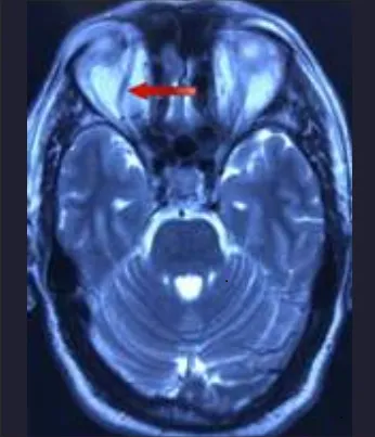
Clinical image showing chemosis, conjunctival injection, and slight proptosis of the right eye. Fundoscopy demonstrated papilledema and retinal venous engorgement of the right eye. Based on the above clinical presentation and ocular findings, a presumptive diagnosis of a CCF was made. A magnetic resonance imaging (MRI) of the brain was ordered which revealed marked enlargement of the right superior ophthalmic vein (SOV). The patient was then transferred to the department of neurovascular intervention for further management.
Brain MRI, axial view, demonstrating a dilated superior ophthalmic vein (red arrow)
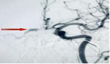
A digital subtraction angiography (DSA) was performed that revealed a Type D CCF with an early filing of the right cavernous sinus in the arterial phase. Based on the rapidly deteriorating clinical picture, shunt anatomy, and hemodynamics a decision was taken to symbolize the CCF.
DSA cerebral angiogram revealing a CCF with an early filling of the right cavernous sinus in the arterial phase.
Procedure
The patient was placed supine on the angiographic table. The patient was intubated, and the procedure was performed under general anesthesia following strict aseptic protocol, and simultaneous right transfemoral venous and left transfemoral arterial access was taken. A 5F diagnostic catheter from the left groin puncture was placed at the common carotid artery and was navigated from the right transvenous groin access through the inferior vena cava, right atrium, superior vena cava, and the jugular vein. At the level of the jugular sigmoid junction, a microcatheter was maneuvered into the inferior petrosal sinus(IPS) leading to the CS. After cannulating the CS, an angiogram was performed to confirm the position of the microcatheter. Embolization was done with Onyx on the left side and coils on the right side of the CS.
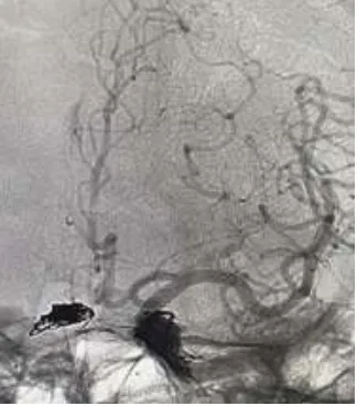
DSA cerebral angiogram showing the placement of coils on the right side and Onyx embolization on the left side.
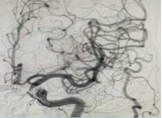
The final angiogram demonstrated occlusion of the CCF with arterial branches within the normal limits. The procedure was completed, and the patient was extubated without any new neurological deficit. A total of five coils and 2ml of Onyx were used to achieve complete embolization
The final check angiogram demonstrating occlusion of the CCF with the normal arterial flow.
There was a significant improvement in the vision, diplopia, and redness in both eyes within 48 hours post procedure. The patient was discharged on the third day postoperatively. At his second-week follow-up, the patient's ocular redness, proptosis, headaches, and visual disturbances had completely resolved.
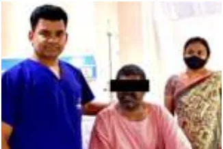
Clinical picture 48 hours postoperatively showing a noticeable reduction of the right eye redness within 48 hours post-procedure

Read this page in:
Feeling unwell?
Book Doctor Appointment in 30 Sec