Arteriovenous malformations in the mediastinum are rare and extremely unusual in the middle mediastinum. The present report describes a 26-year male with spontaneous massive hemoptysis due to arteriovenous malformation in the middle mediastinum and successful management by endovascular embolization. Spontaneous massive hemoptysis can be a presenting feature of arteriovenous malformation in the middle mediastinum and endovascular embolization can be curative.
Case Report
A 26-year male presented with a complaint of spontaneous massive hemoptysis (500-600 mL) and a history of 2 episodes of small quantities (5-15 mL) of hemoptysis in the last 2 weeks. There is no history of fever or prior infections like tuberculosis.or chest trauma. His medical history, physical examination, routine blood tests, and electrocardiogram were unremarkable.
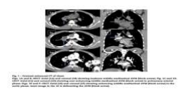
A chest radiograph revealed a mildly bulky right hilum. Non-enhanced computed tomography (CT) of the chest revealed abnormal isodense soft tissue in the middle mediastinum (Figs. 1A and B). CT pulmonary angiogram phase revealed no obvious pulmonary arterial feeders (Figs. 1C and D). Contrast-enhanced CT of the chest in the aortic phase revealed an irregular meshwork of intensely enhancing dilated serpiginous vessels in the middle mediastinum of size 3.5 × 3.0 cm located in the right hilar region, around the right main bronchus (Figs. 1E and F).
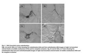
A small component of the meshwork was noted in the subcarinal region. These findings were in favor of AVM. The lesion was enhanced in the aortic phase suggesting its supply from the bronchial artery. So, for better delineation of angioanatomy, digital subtraction angiography (DSA) was considered. A finding of achalasia cardia was noted. DSA of the aorta and selective angiograms of the right intercostobrachial trunk gives rise to the first bronchial artery (Fig. 2A), a second bronchial artery from the aorta (Fig. 2C), left bronchial artery and posterior intercostal arteries confirmed the middle mediastinal AVM supplied by 2 right bronchial arteries and draining into the right superior pulmonary vein (Figs. 2A and C).
The treatment options of surgery and embolization were put forward, out of which the patient gave consent for embolization. Endovascular embolization was done with polyvinyl alcohol particles (PVA-500) and gel foam, after selective cannulation of the first bronchial arterial feeder (Figs. 2A and B) and second bronchial arterial feeder (Figs. 2C and D). With the complete exclusion of the AVM from the circulation at the end of the procedure with no complications. The patient was discharged and is on regular follow-up for 4 years with no recurrence of hemoptysis.
Discussion
Arteriovenous malformations (AVM) of the mediastinum are extremely rare [1–8] lesions and are more commonly present in childhood. This vascular anomaly was first described in the mediastinum by Lunde and colleagues in 1984 [6]. Since then, fewer than 10 cases of posterior mediastinal AVMs, 2 cases, and 1 case of anterior and middle mediastinal AVMs in adults have been documented [8]. To the best of our knowledge from the literature, symptomatic middle mediastinal AVM with massive hemoptysis in an adult is extremely rare.
AVMs are reported in many organ systems of the body. In the thorax, pulmonary AVMs are more commonly encountered and may be associated with hereditary hemorrhagic telangiectasia. (Osler-weber-rendu disease). Our case of middle mediastinal AVM is extremely rare and was supplied by 2 right bronchial arteries with no association with Osler-weber-rendu disease. An AVM may be asymptomatic and becomes symptomatic if it gets infected or enlarges and exerts pressure on vital mediastinal structures, such as the trachea or superior vena cava
Pneumonia VAP with ARDS In view of prolonged ventilation, percutaneous tracheostomy was done. Gradually patient started improving, supports were weaned off, and decannulation was done on the post- operative day-17. Post-decannulation, the patient remained stable, had no new clinical deterioration, and was discharged on the 27th day of admission with the advice to follow up for a zygomatic implant and nasolabial flap to cover the palate.Severe hemorrhage is also a risk, although, to date, only 1 patient with spontaneous rupture of posterior mediastinal AVM with bilateral hemothorax has been documented [4].
Our case of middle mediastinal AVM with spontaneous rupture into the airway with massive hemoptysis is unusual and hasn’t been addressed. Asymptomatic AVMs may be managed conservatively, using a wait and watch approach. If the AVM is symptomatic as was our case, treatment options are embolization or surgery or preoperative embolization followed by surgery.
Complete surgical removal of mediastinal AVMs may be complicated by encroachment and adherence of the lesion on adjacent vital mediastinal structures such as major airways and great vessels [8]. In our case, there was encroachment of the right intermediate bronchus, so surgical resection was thought to be complicated and not feasible. The patient consented to endovascular embolization, which was carried out successfully with complete exclusion of middle mediastinal AVM from the circulation. There was no recurrence of hemoptysis on follows up.
Conclusion
Arteriovenous malformation in the middle mediastinum of an adult presenting with spontaneous hemoptysis is extremely rare and endovascular embolization can be curative. To the best of our knowledge from the literature, such a case is extremely rare.
Contributors
Senior consultant Interventional Radiologist
Consultant Interventional Radiologist
News Letter
Medicover Hospitals Impact Newsletter August 2022